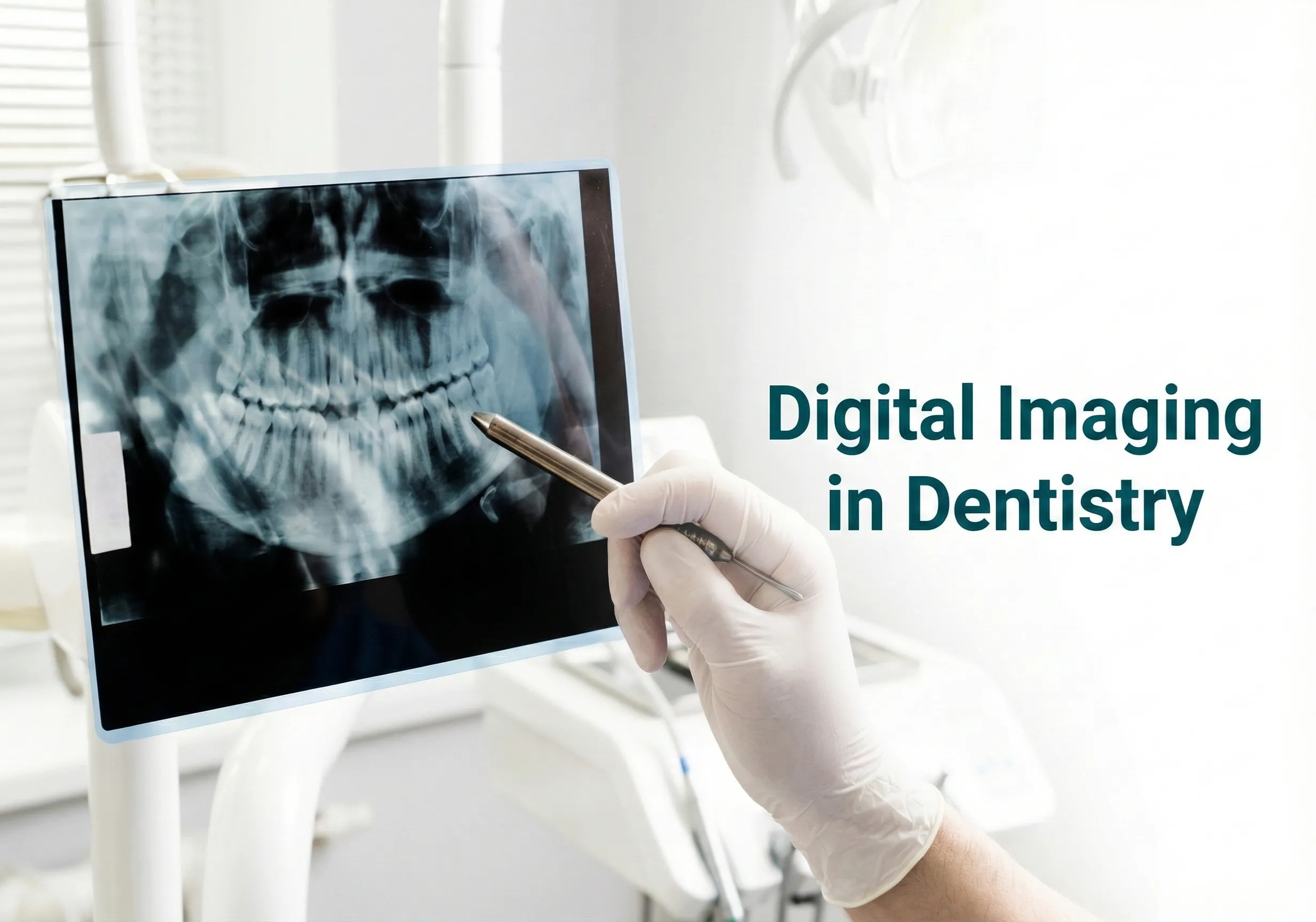
At Todays Dental Lexington, accurate diagnosis is the foundation of effective dental care. Digital imaging is a modern method of dental radiography that has transformed how dentists diagnose oral conditions.
By replacing traditional film-based X-rays, digital radiography allows faster, clearer, and more efficient imaging—benefiting both patients and clinicians.
Dental imaging uses X-rays to capture detailed images of teeth, bones, and surrounding structures.
Many dental conditions—such as cavities, periodontal disease, or bone loss—are difficult to detect through clinical examination alone.
Digital X-ray images allow dentists to identify these conditions early, saving time and improving treatment planning.
At Todays Dental Lexington, digital imaging helps us diagnose conditions accurately and explain treatment needs clearly to our patients.
X-rays were first discovered in 1895, and conventional film radiography was widely used for decades.
Since the mid-1980s, digital radiography has largely replaced traditional screen-film methods.
Digital imaging uses solid-state sensors to capture images, which are instantly displayed and stored on a computer.
This advancement has significantly improved diagnostic precision and workflow efficiency.
Clinical advantages include:
Digital imaging is also widely used in forensic dentistry for age estimation, dental identification, postmortem examinations, and legal documentation.
Intraoral radiographs are taken from inside the mouth and provide detailed views of teeth and supporting structures.
Common intraoral techniques include:
Extraoral radiographs are taken from outside the mouth and are especially useful when mouth opening is restricted due to trauma or pathology.
Common extraoral techniques include:
Digital radiography requires significantly less radiation exposure compared to traditional E-speed film.
One digital image can be adjusted for multiple diagnostic purposes—lighter images for bone evaluation and darker images for caries detection—without retaking X-rays.
This makes digital imaging both safe and efficient for patients.
At Todays Dental Lexington, we use digital imaging to ensure precise diagnosis, safer imaging practices, and transparent communication with our patients.
Advanced imaging allows us to detect problems early and tailor treatment plans effectively.
If you need an accurate dental diagnosis or want to understand your oral health better, book a checkup at our Lexington office today — routine checkups include digital X-rays when needed.
We’re here to help you make informed decisions about your dental health.
Digital imaging replaces photographic film with electronic sensors that send an image straight to a computer. It covers intraoral and panoramic radiographs, cone beam scans, and intraoral cameras. The images appear within seconds, can be magnified and adjusted for contrast, and are stored in the patient record so changes are easy to compare over time.
Digital sensors need considerably less radiation than traditional film to produce a usable image, so the already small dose from dental radiographs is reduced further. Lead aprons and thyroid collars add protection, and dentists follow the principle of taking only the images that will genuinely change a diagnosis or a treatment plan.
There is no fixed schedule. The interval depends on individual risk, including decay history, gum condition, age, and any current symptoms. Someone with frequent cavities may need images more often than a patient with stable oral health. A dentist decides based on clinical examination rather than taking routine images at every visit.
Radiographs reveal what is hidden below the surface, such as decay between teeth or under existing fillings, infection at the tip of a root, bone loss from gum disease, cysts, impacted teeth, and the position of nerves. Three dimensional scans also help plan implants and complex extractions with far greater accuracy.
Tell us a little about what you need — we'll be in touch shortly.
458 Old Cherokee Rd suite 100, Lexington, SC 29072, United States
Hi! I'm Tooth fairy! Your AI Assistant Ask me anything